Bone Cross Section Slide Labeled : 14 poonam bones
Bone Cross Section Slide Labeled : 14 poonam bones. Related posts of bone cross section labeled bone test anatomy and physiology. We have added a dotted line around the outside of the osteon in case you had trouble picking them out on the previous image. Bone matrix and cells bone matrix osseous tissue is a connective tissue and like all connective tissues contains relatively few cells and large amounts of extracellular matrix. When autocomplete results are available use up and down arrows to review and enter to select. Compare diameters of these cells with those in the next two slides, which are at.
Browse 4,294 bone cross section stock photos and images available, or search for human bone cross section to find more great stock photos and pictures. There is a printable worksheet available for download here so you can take the quiz with pen and paper. Bone in arm pictures 12 photos of the bone in arm pictures bone cancer arm pictures, pictures of bone cancer in arm, bone, bone cancer arm pictures, pictures of bone cancer in arm That's why the color looks different. Microscopic structure of compact bone consists of multiple cylindrical structural units known as osteons or haversian systems.
kidney-slide-labelled-histology | SchoolWorkHelper from swh-826d.kxcdn.com Touch device users, explore by touch or with swipe gestures. We have added a dotted line around the outside of the osteon in case you had trouble picking them out on the previous image. .and hyaline cartilage slides, b) describe the differences you observed between the elastic cartilage and hyaline cartilage slides palaglapu styles data table 3. When autocomplete results are available use up and down arrows to review and enter to select. Smartdraw includes 1000s of professional healthcare and anatomy chart templates that you can modify and make your own. Histology spinal cord and ganglion : Microscope slide showing a cross section of mammalian compact bone. There are two slides to examine.
Mammal compact bone slide, ground c.s.
Related posts of bone cross section labeled bone test anatomy and physiology. 400x this image is from a different slide than the other two images on this page. Same bone section (slide 7) at higher magnification. Very inneficient way to merge verticles. It can be found under the periosteum and in the diaphyses of long bones, where it provides support and protection. The two remodeling sites are in a bone formation phase with osteoid seams (solid arrow) and osteoblasts (open arrows) clearly visible. Before placing your slide on the microscope stage, remember to read the label, examine the slide with your eye and note any visible macroscopic features that might help your examination. Notice the layered effect in the matrix. Consistency in fascicular organization of tibial nerve. Bone cross section slide labeled : Fetal leg, cross section, h&e, 40x (bone marrow in tibia and fibula, developing blood cells, sinusoids, megakaryocytes). .and hyaline cartilage slides, b) describe the differences you observed between the elastic cartilage and hyaline cartilage slides palaglapu styles data table 3. Bone matrix and cells bone matrix osseous tissue is a connective tissue and like all connective tissues contains relatively few cells and large amounts of extracellular matrix.
Bone, ground thin, human, longitudinal section digitalscope compare these two slides with each other and with webslide 74 identify in each slide: Nuclei are central but appear only when the section goes through the widest part of the cell. It can be found under the periosteum and in the diaphyses of long bones, where it provides support and protection. Compare diameters of these cells with those in the next two slides, which are at. There is a printable worksheet available for download here so you can take the quiz with pen and paper.
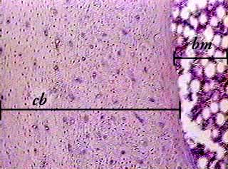
Bone, compact, decalcified c.s. from www.austincc.edu In this image the bar indicates the location of decalcified compact bone. Intervertebral disc, h&e, 40x (bone marrow in spongy bone of vertebrae) virtual slide. Bone matrix and cells bone matrix osseous tissue is a connective tissue and like all connective tissues contains relatively few cells and large amounts of extracellular matrix. The two remodeling sites are in a bone formation phase with osteoid seams (solid arrow) and osteoblasts (open arrows) clearly visible. Bone cross section slide labeled : A long bone has two parts: We have added a dotted line around the outside of the osteon in case you had trouble picking them out on the previous image. Nuclei are central but appear only when the section goes through the widest part of the cell.
That's why the color looks different.
Bone in arm pictures 12 photos of the bone in arm pictures bone cancer arm pictures, pictures of bone cancer in arm, bone, bone cancer arm pictures, pictures of bone cancer in arm Notice the layered effect in the matrix. 400x this image is from a different slide than the other two images on this page. Smartdraw includes 1000s of professional healthcare and anatomy chart templates that you can modify and make your own. It can be found under the periosteum and in the diaphyses of long bones, where it provides support and protection. Using clear epox glue, bind the section to the microscope glass slide. Compact bone and spongy bone: Related posts of bone cross section labeled bone test anatomy and physiology. At this level of magnification, the fundamental structure of compact bone is visible. The osteocytes are arranged in concentric rings of bone matrix called lamellae (little plates), and their processes run in interconnecting canaliculi. In the center of each osteon is the central canal, a space that houses blood vessels and nerves that supply bone. Bone cross section slide labeled : Before placing your slide on the microscope stage, remember to read the label, examine the slide with your eye and note any visible macroscopic features that might help your examination.
Compare diameters of these cells with those in the next two slides, which are at. In this image the bar indicates the location of decalcified compact bone. Notice the layered effect in the matrix. Bone in arm pictures 12 photos of the bone in arm pictures bone cancer arm pictures, pictures of bone cancer in arm, bone, bone cancer arm pictures, pictures of bone cancer in arm 400x this image is from a different slide than the other two images on this page.
Compact Bone | ClipArt ETC from etc.usf.edu That's why the color looks different. Bone matrix and cells bone matrix osseous tissue is a connective tissue and like all connective tissues contains relatively few cells and large amounts of extracellular matrix. In the center of each osteon is the central canal, a space that houses blood vessels and nerves that supply bone. The outlined area is a cross section of an osteon of compact bone. In this image the bar indicates the location of decalcified compact bone. Compact bone is the denser, stronger of the two types of bone tissue ( (figure) ). This slide contains a section of dried compact bone. Related posts of cross section of human bone diagram bone in arm pictures.
In this image the bar indicates the location of decalcified compact bone.
Free online quiz compact bone microscope slide labeled. .and hyaline cartilage slides, b) describe the differences you observed between the elastic cartilage and hyaline cartilage slides palaglapu styles data table 3. Cross section of a bone diagram : Compact bone and spongy bone: *none of the slide images above are shown at their actual scale. The outlined area is a cross section of an osteon of compact bone. The diaphysis and the epiphysis. Cut the section to dimensions of about 5mm by 5mm chip. The structure of a long bone allows for the best visualization of all of the parts of a bone ( figure 6.7 ). The basic unit of structure in compact bone is the osteon. Try pressing the section on the slide to ensure that the layer of glue is as thin as possible. Browse 4,294 bone cross section stock photos and images available, or search for human bone cross section to find more great stock photos and pictures. Bone in arm pictures 12 photos of the bone in arm pictures bone cancer arm pictures, pictures of bone cancer in arm, bone, bone cancer arm pictures, pictures of bone cancer in arm
Microscopic structure of compact bone consists of multiple cylindrical structural units known as osteons or haversian systems bone cross section. Before placing your slide on the microscope stage, remember to read the label, examine the slide with your eye and note any visible macroscopic features that might help your examination.
Hot Air Balloon Crash Texas / Texas Hot Air Balloon Crash No Survivors Among 16 On Board Bbc News . Here's the latest information on the crash: Texas hot air balloon crash: It is the deadliest hot air balloon crash in the us. Mr rowan had just started a job as chief of clinical trials in burns and trauma at the us army institute of surgical research. With 16 people on board flying in the summertime maybe there wasn't enough lift. More than 150 commercial hot air balloon companies are in operation in north america, he said. A hot air balloon with 16 people on board caught fire and crashed in central texas. The deadliest hot air balloon accident in us history occurred in texas in 2016. Texas hot air balloon crash: On july 30, 2016, sixteen people were killed when the hot air balloon they were riding in struck power lines, crashed and caught fire in the unincorporated community of maxwell, near lockhart, texas, 30 miles (50 km) south of the state capital austin.
Cara Mengaktifkan Data Modem / Cara Mengaktifkan Jaringan Data GSM Pada Smartfren ... . Mengaktifkan idm di browser beda dengan aktivasi loh ya. Berikut cara singkat dan paling mudah untuk mengaktifkan atau manyalakan fitur 4g pada smartphone android. Untuk aktivasi, salah satu caranya menggunakan serial number idm. Untuk mencegah terjadinya factory reset yang mengharuskan kita menghapus semua data dan aplikasi yang tersimpan di hp samsung, kami sarankan untuk mencoba cara menonaktifkan mode aman samsung berikut ini. Untuk mengaktifkan data roaming ini kamu harus pikir dua kali untuk mengaktifkan. Selamat berselancar di internet, dan sekian postingan tentang cara mengaktifkan modem zte, setting atau. Artikel ini menjelaskan cara mengkonfigurasi mode tablet agar pc dapat beralih antara mode tablet dan desktop baik secara manual atau otomatis. Ternyata responnya banyak sekali, karena pilih lan host kemudian centang lan 1 dan 2 untuk mengaktifkan port lan 1 dan 2 agar m
Un ego parfois trop amplifié ne saurait qu'éloigner le franc maçon de sa raison d'être initiale. Par contre c'est un ego démesuré qui nous empêcherait d'accepter les autres, et de se soumettre aux principes de la démocratie. Bien au contraire, il nous donne la motivation pour mener nos combats contre les injustices, contre le fanatisme, contre les préjuges, etc. Dans une lettre qu'il adressa le 2 mars 1763 à chaillon de jonville, substitut général du grand maître de la grande loge de france, il fit suivre sa signature des titres suivants: Maître écossais, g(rand) a(rchitecte), r(oï)al arch, chevalier d'orient, d'occident, du soleil, de l'aigle noir, r(ose) c(roix), g.i.g.e.ch.k. Telecharger Lettre De Motivation Gratuite En Pdf â€" Lettre from ltmodele.urez.us (c'est à dire grand inspecteur, grand el
Does Dr Jeff Live Above His Animal Hospital Our Fearless Leader – Dr. Jeff Young Dr. Jeffrey Young graduated from the Colorado Country University, School of Veterinary Medicine in 1989. Dr. Immature believes his humane ethics come from working every bit an Fauna Control Officeholder during his university training, where he witnessed corruption and neglect of healthy companion animals. He established Planned Pethood Plus, Inc. (PPP) in 1990 as a low-cost spay/neuter dispensary and now provides affordable full-service veterinary care. Planned Pethood is well known for depression-cost mobile neutering clinics, Native American Reservation piece of work and preparation of veterinarians from around the earth in more efficient surgical technique. Dr. Young has served on numerous Humane Social club boards and has advised mobile surgical units all across America. In the year 2000, Dr. Immature founded Planned Pet


Comments
Post a Comment